NONINVASIVE OPTICAL AND LASER
MEDICAL DIAGNOSTICS
(noninvasive clinical spectrophotometry, bio-spectrophotometry)
The analysis of different tendencies in a modern medical diagnostics and a treatment shows that today in the overwhelming majority of the cases those methods come to the first place that minimize invasiveness, pharmaceutical (chemical) and other ecologically and physiologically undesirable effects on the human’s organism. Besides that, the increase of human population and increase of its sickness rate throughout the World increases a load on a medical staff in clinics and puts in the forefront those medical technologies that produce a maximal effect at a minimum waste of time. These requirements are in full measure met by the latest optical (laser) clinical noninvasive diagnostic methods that recently began to appear in many leading countries all over the World, including our Russia. The laser methods of diagnostics are based on a simple, obviously fact, that the total optical properties of biological soft tissues always are a function of physiological, functional and pathology state (condition) of tissues and organs. Different spectral optical properties of human tissues and liquids, especially blood, are different for normal and pathological cases, so there is a possibility to determine the tissues' and organs' clinical state by means of measuring "in vivo" ("in situ") its spectral optical properties - absorbance, scattering, reflectance, fluorescence, etc. Strictly speaking, there are the same base physical principles in noninvasive optical diagnostics that are used in a conventional laboratory spectroscopy technique in conventional clinical tests excepting the advantages of "in vivo" and "real-time" operating mode. So, the most of noninvasive optical and laser medical diagnostic methods and techniques (excepting laser tomography) in full manner could be called together today in a total as the "noninvasive clinical spectrophotometry technique".
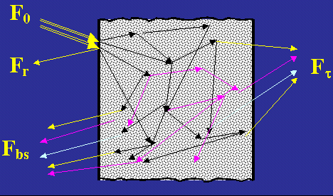
To realize the optical noninvasive diagnostic technique, a tested area of tissue is illuminated by low-intensive light flux (F0) of different waveband and the backscattered (Fbs) and/or transmitted by the tissue light fluxes (Fig.1) are registered by a photodetector. Thus, the first step of any spectroscopy diagnostic methods is the registration of the secondary optical radiation after its interaction with biotissue.
Fig. 1.
It is important to understand better the specialties of optical noninvasive technique - the right understanding that in a tissue there take place a lot of linear and nonlinear physical phenomena of light-tissue interaction: light absorption and scattering, fluorescence, Doppler frequency shift on a moving blood cells, etc., so the spectral and spatial (per a tissue surface) distribution of a registered radiation in comparison with the initial one contain a lot information about the tissue biochemical and/or structure composition. To extract this information on the next step of diagnostic data processing the electrical signal, proportioning to the registered spectral light intensity, is directed to a processing computer to calculate the existing tissues’ optical properties - the optical transport coefficients in accordance with the applied physical and mathematical model of the general theory of light transport and scattering in a tissue (see, for example, mathematical models of linear interaction of laser radiation with different media). Here we have to specially note that it is not a direct optical properties measurement. It is the so-called indirect technique that requires some next calculation routines - the so-called "inverse optical task algorithms". Today in the most majority of cases the general Light Transport Theory is usually used to calculate optical transport coefficients for the tested biological media. A number of noninvasive technique (photoplethismography, tissues reflectance oximetry or laser Doppler flowmetry, for example) use the additional spectral analysis algorithms to determine dynamic parameters and rhythms of the registered optical signals.
But any optical properties of tissue or blood are not quite medical or biological information about a patient yet. What follows for a doctor from the fact, for example, that the patient's skin has a transport scattering coefficient 0.5 cm-1 at a wavelength 632 nm? Nothing. The same question can be asked when the intensity of fluorescence only is presented for a doctor as the final information of the fluorescent diagnostic procedure. Shortly speaking, this level of information is not a final medical level of diagnostic information. It is only a physical data level. The medical and biological levels of information will appear when, on the basis of measured physical data, the information of biochemical molecules content in a tissue or blood will be revealed. For example, in tissues oximetry the oxygen saturation in the blood, i.e. the content of the oxygenated fraction of the hemoglobin in blood is calculated and that is the final medical information level. Absorption spectroscopy can produce the information about content of water, lipids, melanin, etc. in a tissue. Scattering spectroscopy and tomography techniques give the information about a morphological structure of the tissue. Different dynamic methods (Doppler flowmetry, plethismography) calculate the dynamic parameters of the blood flow. And so on. All this calculation procedures must be presented in a system's computer software as well.
For a medical user a laser noninvasive diagnostic procedure consists
only in bringing of optical fiber or another optical probe to a slight contact
with biotissue (Fig.2) and reading the final medical results of
diagnostic procedure in a real time mode from the computer's monitor.
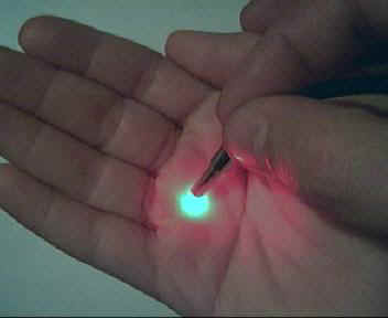
Fig.2.
Today there are in the World a number of promising noninvasive optical and laser diagnostic methods, which are a subject of intensive scientific research at a present: the fluorescent diagnostics, elastic scattering spectroscopy, laser Doppler flowmetry, laser coherent and diffusion tomography, tissues reflectance oximetry, pulsoximetry, etc. A number of non-invasive optical diagnostic apparatus are already widely presented on the medical equipment’s market (puls-oximetry and laser Doppler flowmetry systems, for example). Another series of apparatus are now on a final stage of the way to the market (fluorescent systems, for instance). In different expert’s opinion, it takes not a long time to develop this “scientific level” of technology to a "commercial level" of technology and equipments. Moreover, today it is already evident, that the noninvasive optical diagnostics can become a new, separated and widespread medical technology which can be very effective in different areas of medicine: surgery, oncology, urology, dermatology, gynecology and others. It can give a lot of additional information about relevant content in a tissue a number of important biochemical components (hemoglobin fractions, porphyrin, NADH, etc.), about functional activity of that in a tissue, about blood supply for a tested tissue, etc. These techniques can be successfully used in different brunches of modern clinical medicine (angiology, dermatology, oncology, etc.), in a sport medicine, in a doctor’s training process, student education, so the business and practical perspectives of that are very interesting.
For more information see a paper D.A.Rogatkin, L.G.Lapaeva "Prospects for development of noninvasive spectrophotometric medical diagnosis" // Biomedical Engineering, v.37, No.4, 2003. - pp.217-222.
Have a look at this paper in PDF (116KB)